Thyroid imaging tests
Thyroid imaging tests are often used to diagnose thyroid disease. This page describes the thyroid imaging tests that are used for the diagnosis and management of thyroid disease
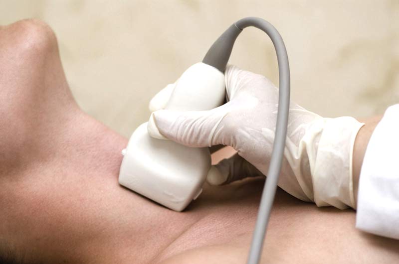
Thyroid ultrasound
- This simple test is usually the first thyroid imaging test requested and uses sound waves to image the thyroid – the sound waves are emitted from a small hand-held transducer that is passed over the thyroid
- A lubricant jelly is placed on the skin so that the sound waves transmit more easily through the skin and into the thyroid and surrounding structures
- This test is quick, accurate, cheap, painless, and completely safe. It usually takes only about 10 minutes and the results can be known almost immediately thyroid ultrasound will help determine the nature of a thyroid nodule, and therefore if a biopsy is required
Ultrasound characteristics indicating that a thyroid nodule is benign include:
- Sharp edges seen all around the nodule
- A nodule filled with fluid and not live tissue (a cyst)
- Lots of nodules throughout the thyroid (usually a benign multi-nodular goitre)
- No blood flowing through the thyroid nodule (not live tissue, likely a cyst)
A cystic thyroid nodule is often benign but larger thyroid cysts have an greater risk of associated cancer – once a thyroid cyst reaches 4cm or more in diameter the risk or cancer is around 20%
Thyroid ultrasound is also capable of identifying thyroid nodules before they can be felt and can be used to help guide and increase the accuracy of thyroid biopsy
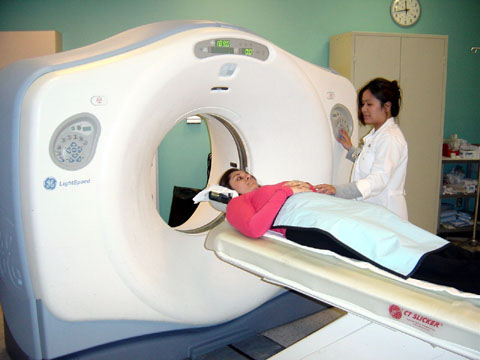
CT Scan
CT Scanning is helpful in the evaluation of thyroid disorders where:
- There are pressure related symptoms
- The thyroid is growing into the chest – retrosternal goitres
- For the evaluation of patients with thyroid cancer
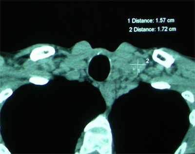
Thyroid Gland CT Scanning and CT scan image showing a thyroid tumour:
MRI scan
MRI scanning is done when the size and shape of the thyroid needs to be evaluated – MRI may be recommended instead of a CT scan because it doesn’t require any injection of contrast dye, and doesn’t require radiation
Radioactive Iodine Uptake (RAI-U) and thyroid isotope scan
Thyroid scan
- Radioactive iodine uptake is a way of measuring thyroid function – by measuring how much iodine is taken up by the thyroid gland (RAI uptake)
- Thyroid cells normally absorb iodine from our blood stream (obtained from foods we eat) and use it to make thyroid hormone
- In this test, a small dose of an isotope, usually radioactive iodine 123, is given in pill form and several hours later, the amount of iodine in the bloodstream is measured and the amount of iodine that goes into the thyroid gland can be measured as the thyroid uptake
- A radioactive iodine uptake (RAI-U) test can help tell whether a person has Graves’ disease, toxic multinodular goiter, or thyroiditis
- Thyroid isotope scanning tests how well the thyroid gland is functioning and requires giving a radioisotope to the patient and letting the thyroid gland concentrate the isotope (just like the iodine uptake scan above)
- Therefore thyroid isotope scanning is usually done at the same time that the iodine uptake test is performed – pictures of the thyroid gland are taken with a special camera after the injection of a radioactive iodine or technetium isotope
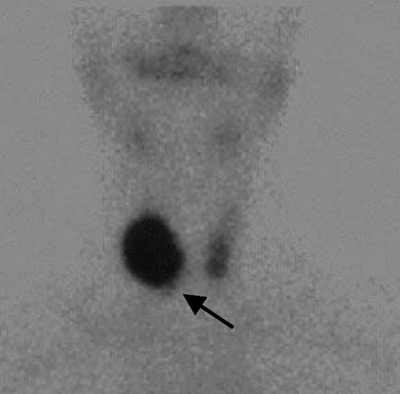
- The picture taken with thyroid isotope scanning shows how well the thyroid gland is functioning, and in particular, whether a thyroid nodule is functioning
- Thyroid isotope scanning allows the classification of thyroid nodules as non-functioning (cold thyroid nodules), normally functioning (warm thyroid nodules) and hyperfunctioning (hot thyroid nodules)
- Thyroid isotope scanning is helpful for patients with thyroid nodules in the presence of a low TSH level as hot thyroid nodules are usually benign
- Overall 80% of nodules scanned are cold but only 20% of these will be malignant
If you have any questions about thyroid disease diagnosis, thyroid imaging or thyroid or parathyroid surgery, contact your local doctor, who will arrange for you to see a thyroid surgeon.


